
Diagnosis
Diagnosis is the foundation of effective cardiac care.
Before any treatment begins, we need to understand exactly what’s happening, which means identifying exactly which part of the heart is affected, how it’s functioning, and what that means for your pet’s health.
Cardiac conditions can be complex and often subtle in the early stages. A thorough, specialist-led diagnosis gives us a clear picture of your pet’s condition, helps avoid unnecessary or incorrect treatment, and ensures the best possible plan for moving forward.

What To Expect
At Sawgrass Veterinary Cardiology, we take a gentle, pet-centred approach. Your visit begins with a thorough cardiovascular physical examination and blood pressure measurement, giving us an initial understanding of your pet’s heart and circulatory function.
We take time to discuss your pet’s history and any symptoms you’ve noticed at home, then recommend specific diagnostic tests based on those findings – ensuring we only pursue what’s clinically relevant.
You’re welcome to stay with your pet during all diagnostic tests – except radiographs, which require protective shielding and safety protocols. Our clients often tell us they value how we explain what we’re seeing as we go, so you’re part of the process. We aim to discuss results thoroughly during your visit, so you leave with clarity, confidence, and a clear path forward.

Diagnostic Tools We Use

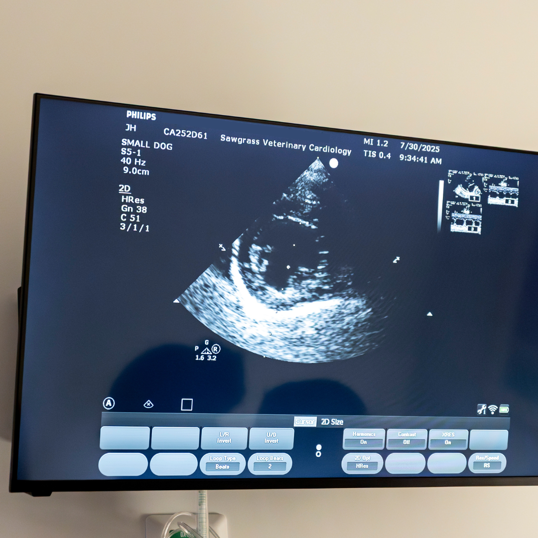
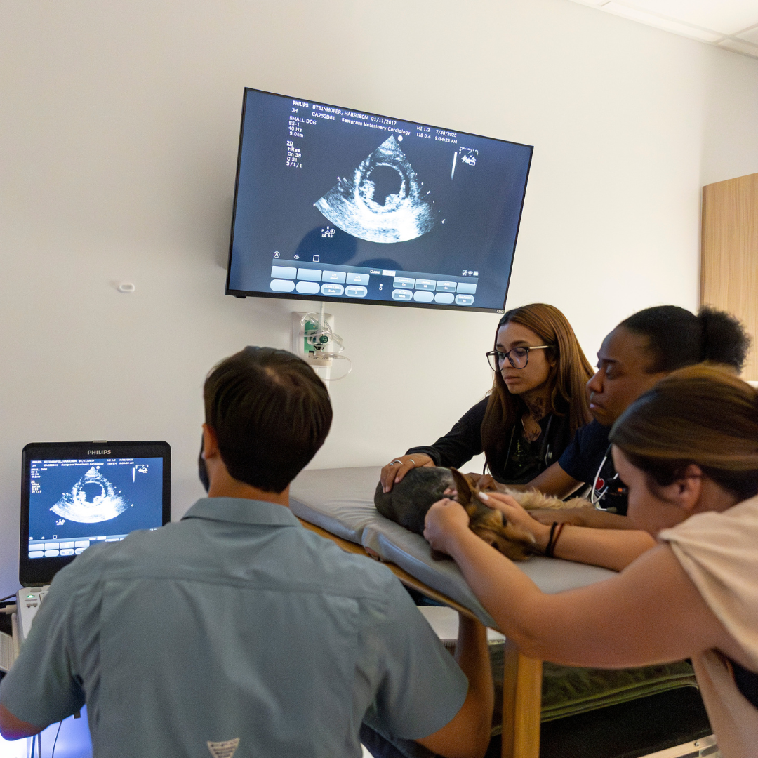
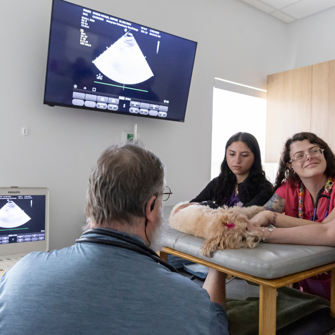
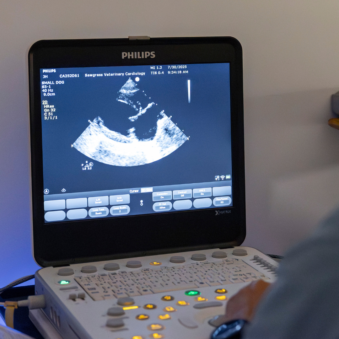
Echocardiography
An echocardiogram is a non-invasive ultrasound of the heart that allows us to see the heart beating in real time. It provides detailed information about heart chamber size, valve function, muscle thickness, and blood flow patterns—all critical for diagnosing conditions like valve disease, cardiomyopathy, congenital defects, or fluid around the heart. Unlike an X-ray, which gives a static image, an echocardiogram shows how the heart functions moment to moment, allowing for more accurate assessment and treatment planning.
At Sawgrass Veterinary Cardiology, we perform all echocardiograms with the pet owner present in the room, so you can be part of the process and see what we see. Most pets do not need sedation and are gently positioned on a padded table while we scan their chest with a specialized probe. The test is painless, typically takes about 10-15 minutes, and results are discussed with you in real time so you leave with a clear understanding of your pet’s heart health and any next steps.
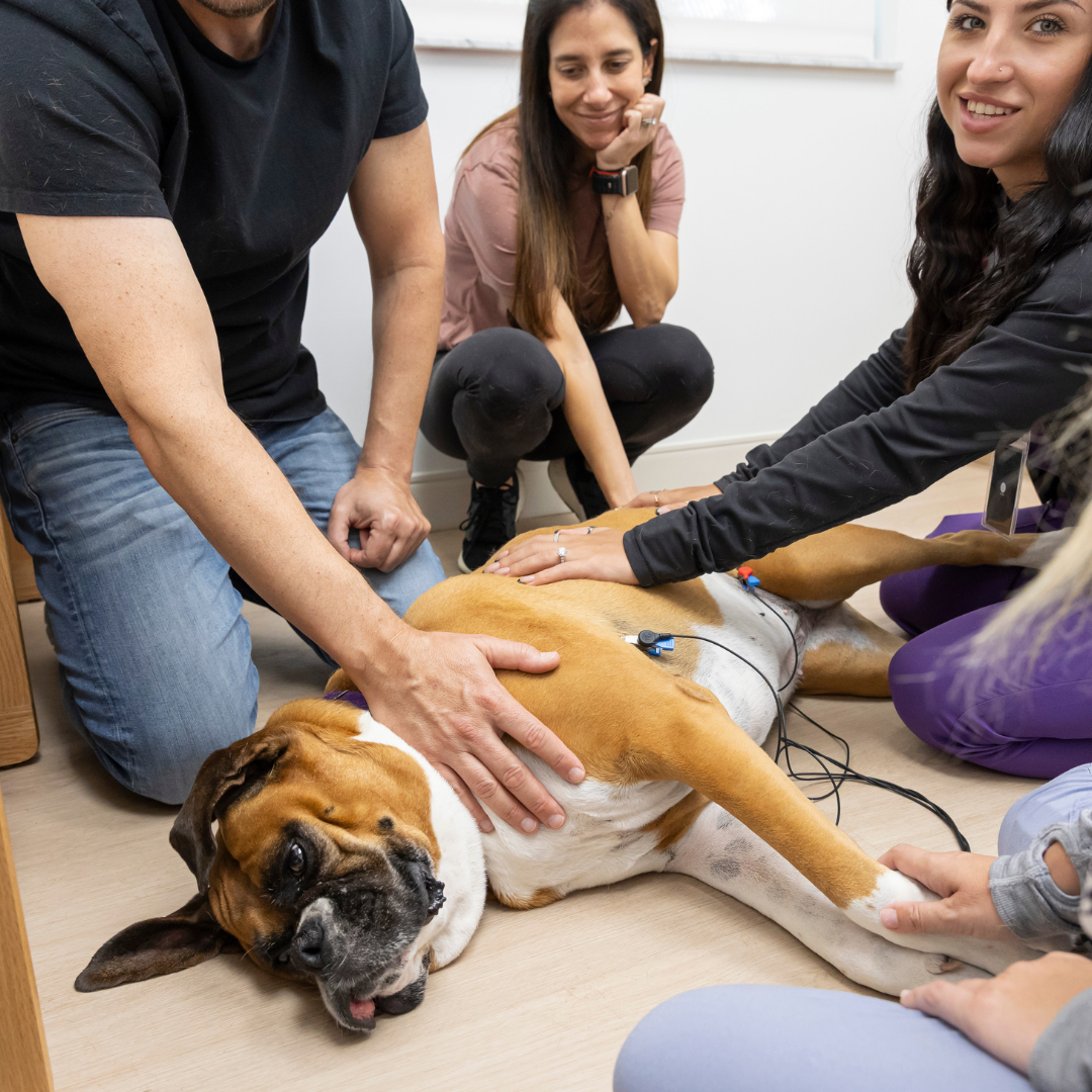
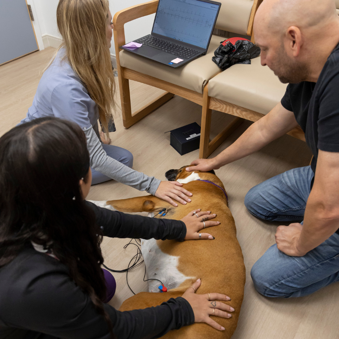
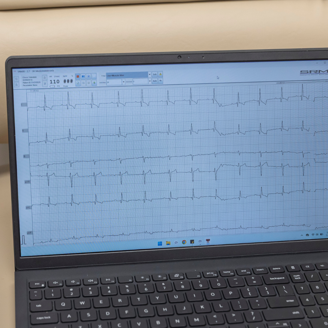
Electrocardiography
An electrocardiogram (ECG or EKG) is a quick, painless test that records the electrical activity of your pet’s heart. It allows us to evaluate your pet’s heart rhythm, rate, and the timing of each heartbeat, helping us diagnose conditions such as arrhythmias, conduction abnormalities, or chamber enlargement. It is often used when pets faint, have irregular heartbeats, or before starting certain heart medications. Even if your pet’s physical exam or chest X-rays appear normal, an ECG may uncover silent electrical disturbances that could be serious if left untreated.
At Sawgrass Veterinary Cardiology, ECGs are performed with the owner present and typically take just a few minutes. Your pet will lie comfortably on their side while small clips are placed on their limbs to record the heart’s electrical signals. Sedation is almost never needed. The results are interpreted in real time by our board-certified cardiologists, and we’ll review the findings with you during your visit. If further evaluation is needed, we may recommend extended monitoring such as a 24-hour Holter monitor, especially in breeds prone to silent arrhythmias like Boxers or Dobermans.
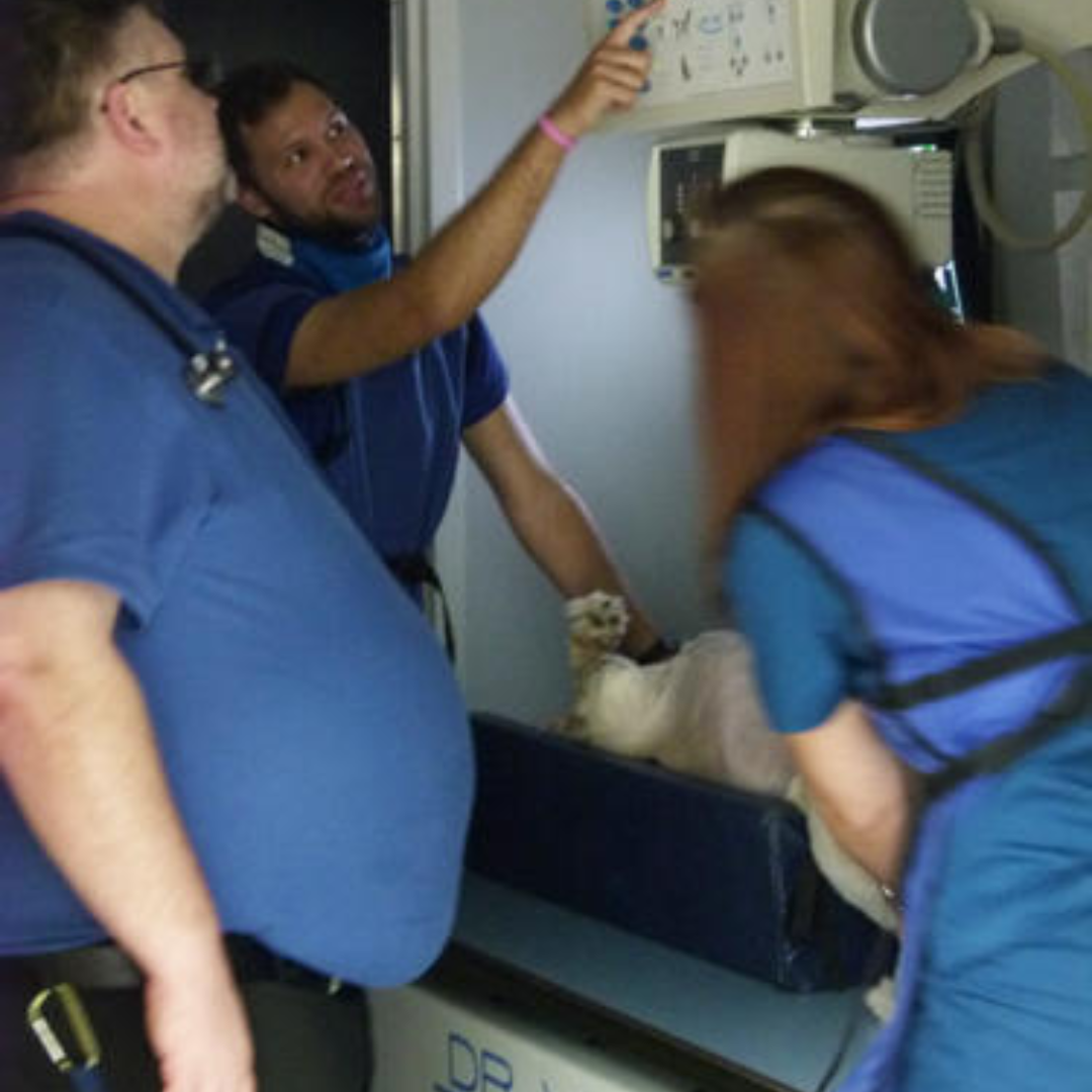
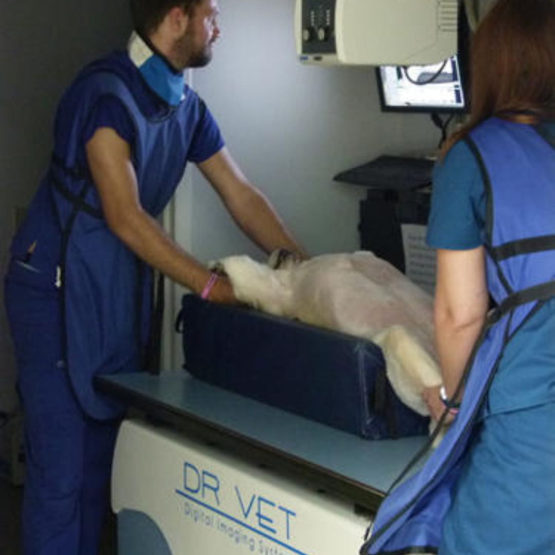
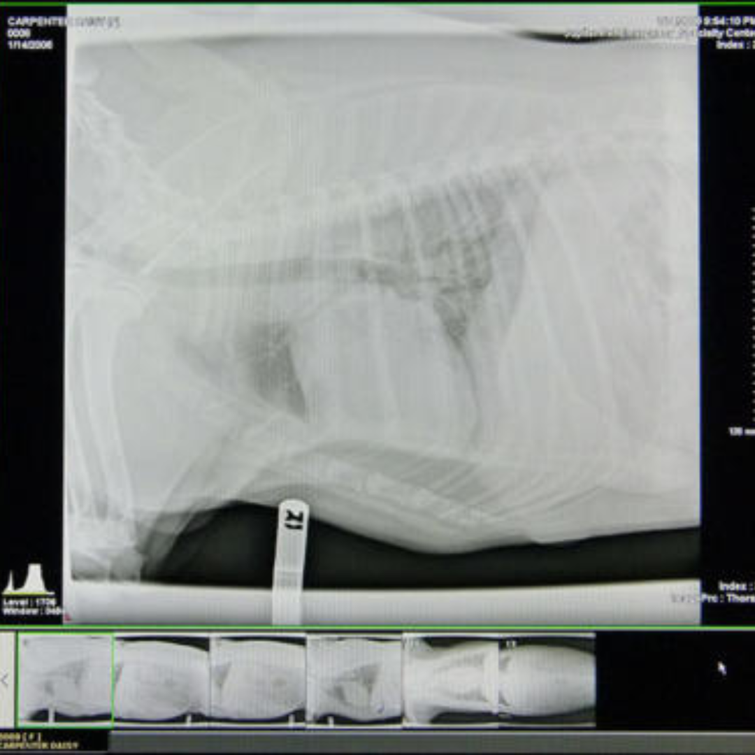
Radiography
Radiography (X-rays) provides a still image of the chest, allowing us to assess the size and shape of the heart, detect fluid in or around the lungs, and evaluate structures like the trachea and major vessels. While it doesn’t show the heart beating like an echocardiogram, radiographs are crucial for identifying signs of congestive heart failure, pulmonary disease, or other conditions that may not be evident on ultrasound alone. It’s an essential piece of the diagnostic puzzle when evaluating heart health.
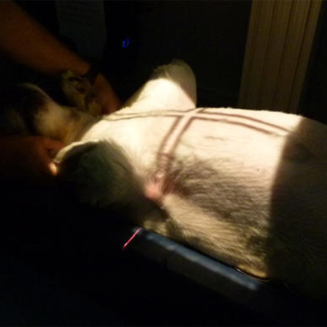
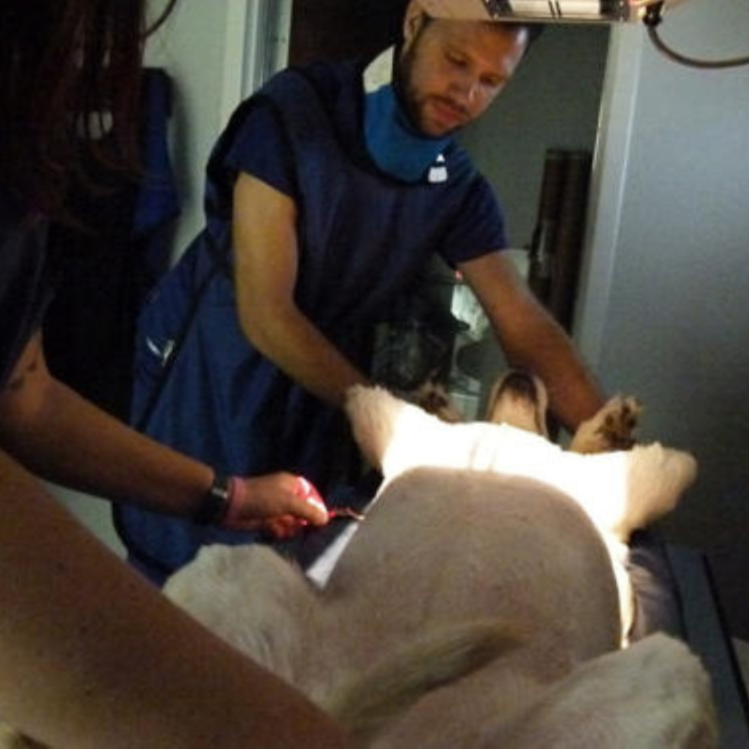
Because radiographs involve exposure to X-rays, we follow strict safety protocols to protect both people and pets. For this reason, pet owners are not able to stay in the room during the imaging process. Our trained team gently positions your pet and takes the necessary images quickly and safely. Sedation is rarely needed. Once the images are taken, we review the findings with you during your appointment and explain how they fit into your pet’s overall cardiac picture.
Advanced Diagnostics
Advanced diagnostics are used selectively to give us a more complete picture of your pet’s heart health when imaging and physical examination alone aren’t enough. These tests help confirm or clarify a diagnosis, monitor progression, or guide treatment decisions in complex cases.
We may recommend:
-
Blood pressure measurement – to assess for hypertension, which can affect or be affected by heart disease.
-
Cardiac biomarkers (e.g. NT-proBNP) – a simple blood test that helps detect hidden heart strain or differentiate cardiac from respiratory causes of symptoms.
-
Holter monitoring – a portable ECG device your pet wears at home to record their heart rhythm over 24+ hours, ideal for capturing intermittent arrhythmias.
-
Genetic testing – available for certain breeds like Boxers to screen for inherited cardiac conditions before signs appear.
-
Fluoroscopy – a dynamic imaging tool (similar to a live X-ray) that may be used in complex interventional or structural cases when available.
Not every pet will need these tests, but when indicated, they provide valuable data that helps us deliver more precise and proactive care. If advanced diagnostics are recommended, we’ll explain why and what to expect, ensuring you’re part of every step.