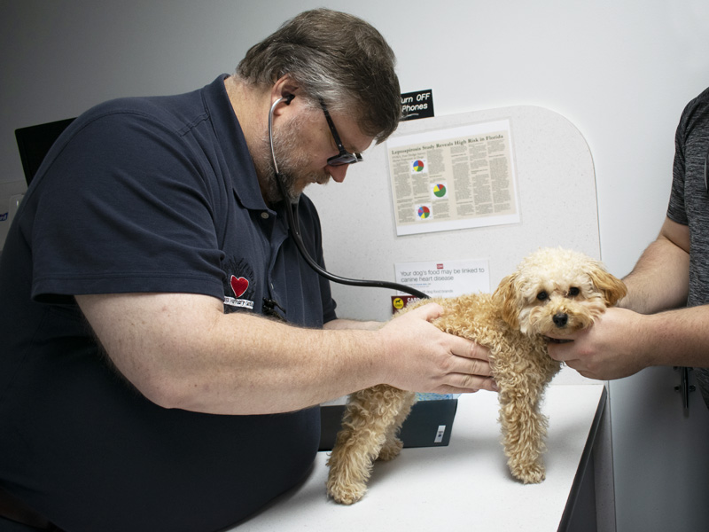
A complete Cardiovascular Physical Examination, including blood pressure analysis, is initially preformed on your pet. This focused examination allows Dr. Carpenter to assess the heart and lungs of your pet. This information is then combined with the history to determine the optimal diagnostic and treatment plan to ensure accurate and cost effective diagnosis of your pets underlying cardiac disease. For more information about what is a heart murmur, please see our blog article.
Further evaluation of a heart murmur by the veterinary cardiologist may include the following:
The cardiologist will carefully listen to the heart with a stethoscope. They will listen to the loudness, location, and timing of the murmur to classify and describe the sound. This will help diagnose the cause of the murmur.
A chest x-ray is a painless test that creates pictures of the structures inside the chest, the heart and lungs. This test is done to help identify potential causes of coughing, decreased exercise ability, difficult or rapid breathing, and episodes of collapse.
An EKG (electrocardiogram) is a simple test that detects and records the electrical activity of the heart. An EKG shows how fast the heart is beating, the heart’s rhythm (steady or irregular), and where in the body the heartbeat is being recorded. An EKG also records the strength and timing of electrical signals as they pass through each part of the heart.
Echocardiography (EK-o-kar-de-OG-ra-fee) is a painless test that uses sound waves to create pictures of the heart. The test gives information about the size and shape of the heart and how well the heart chambers and valves are working. It will often identify the cause of the murmur as well as the overall impact of the murmur on the heart. This allows for the cardiologist to provide treatment recommendations as well as predict the future impact of the heart disease on your pet’s health.