Echo Images Needed to Assess A Patient for TEER Procedure

All content copyright of Colorado State University 2022. Pre-operative evaluation for transapical edge-to-edge mitral valve repair (VClamp device).

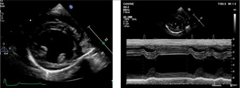
Right parasternal long-axis 4-chamber view

  • 2D (optimized for LA and LV)
  • Color Doppler on MV (include entire LA) and TV
  • Zoomed image of MV
    • 2D
    • Color Doppler on zoomed image

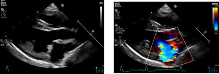
Right parasternal long-axis inflow-outflow view

  • 2D
  • Color Doppler on MV

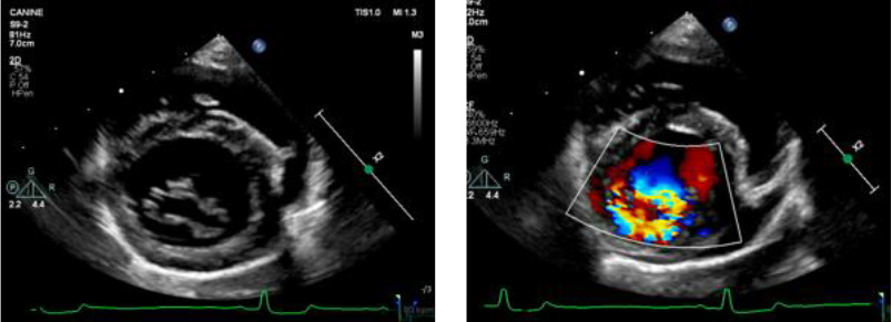
Right parasternal short-axis (LV at level of papillary muscles)

  • 2D
  • LV M-mode

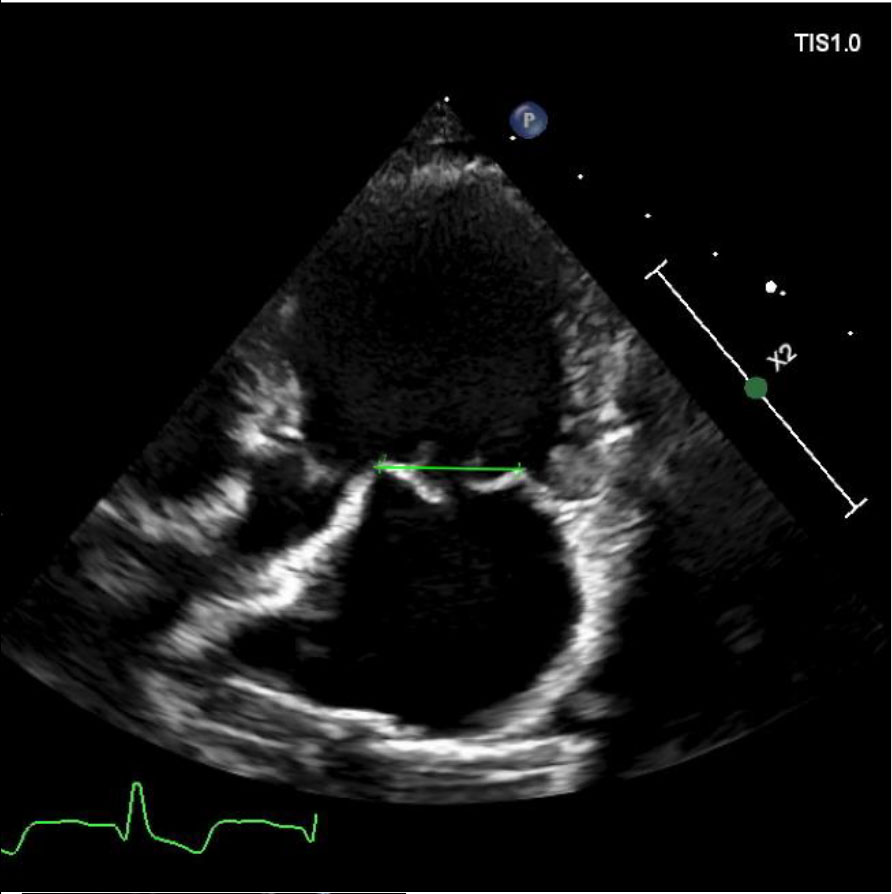
Right parasternal short-axis (LV at level of mitral valve)

  • 2D
  • Color Doppler

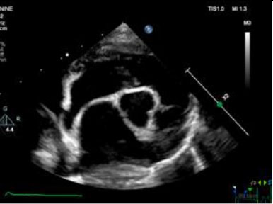
Right parasternal short-axis (heart base – aorta and LA)

  • 2D

Subcostal view

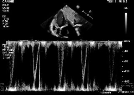
Aortic flow velocity – PW Doppler at valve

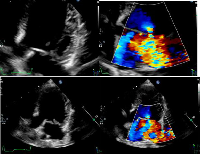
Left parasternal apical four-chamber view

  • 2D
  • MV inflow velocities – PW Doppler
  • Color Doppler on MV and TV
    • MR CW Doppler
    • TR CW Doppler
  • Zoomed image of MV
    • 2D
    • Color Doppler on zoomed image

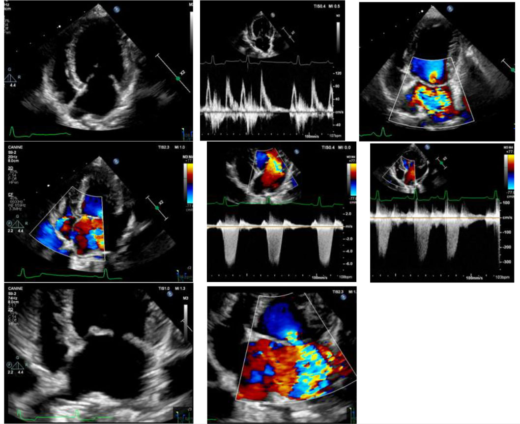
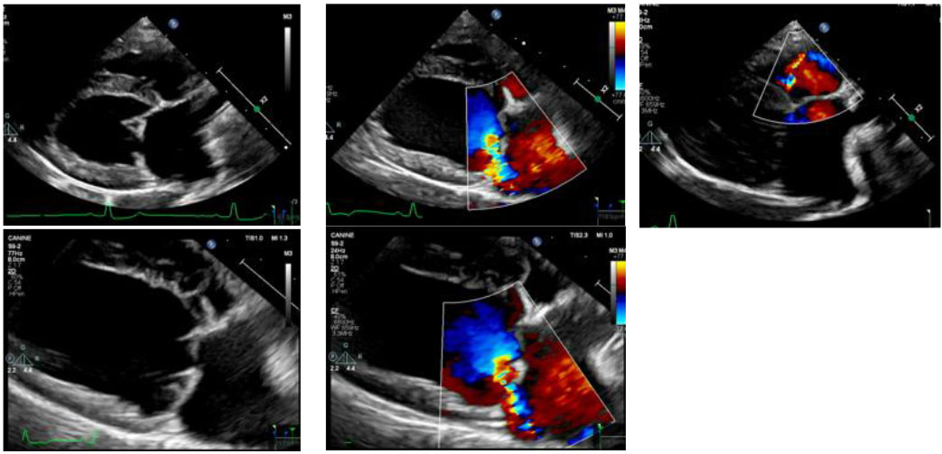
Left parasternal apical five-chamber (inflow/outflow, including aortic valve) view

  • 2D – This view is used to measure the mitral annulus for V Clamp device sizing
  • Color Doppler on MV (include entire LA)
  • Zoomed image of valve
    • 2D
    • Color Doppler on zoomed image

For an Appointment, Please Call 954-487-8357