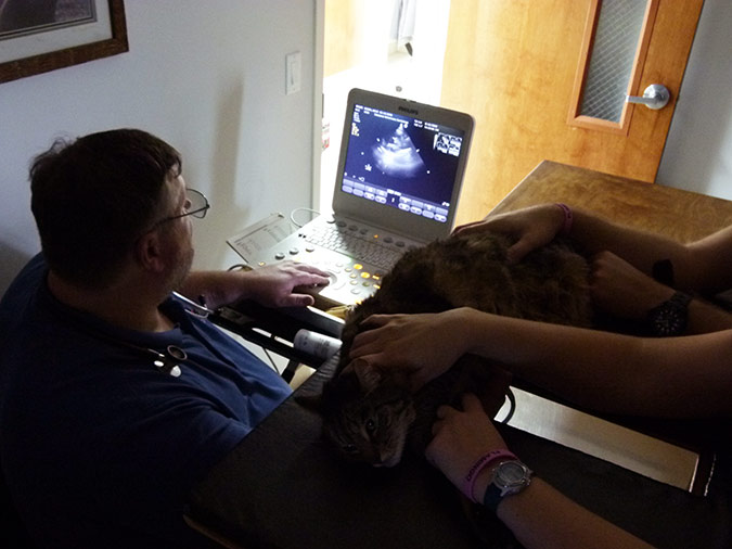
The echocardiogram, also known as cardiac ultrasound, is a non-invasive diagnostic tool with state of the art imagery capabilities for your pet’s heart. It is currently the gold standard of diagnostics tests in veterinary cardiology and uses a probe or transducer to produce and direct sound waves into the body. These waves interact with the tissues in the body and are reflected back to the probe.
By collecting and analyzing these reflected non-harmful sound waves, the ultrasound machine is able to create images of the heart that are then displayed on the monitor. This allows the cardiologist to noninvasively visualize the heart muscle, the heart valves, and the large arteries. Echocardiography also allows us to visualize and measure the manner and speed with which blood flow moves through the heart as well as estimate pressures within the heart.
Patients are not typically shaved for an echocardiogram. Alcohol and ultrasound gel are applied to the fur on each side of the chest as the pet lies on their side. Many measurements are taken during the procedure and analyzed to give Dr. Carpenter immediate results so that a treatment plan can be started immediately. .